This article will break down the key components of your ocular prescription and what they mean for your vision.
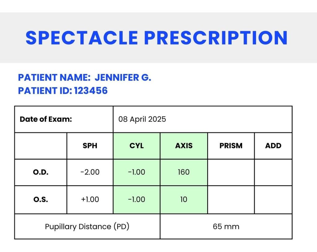
Your prescription typically includes values for each eye, often labelled as OD (oculus dexter, meaning right eye) and OS (oculus sinister, meaning left eye). Sometimes, OU (oculus uterque) may be used if the information applies to both eyes.
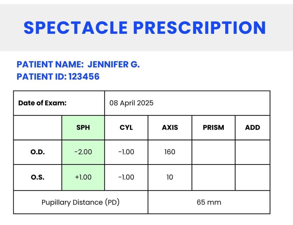
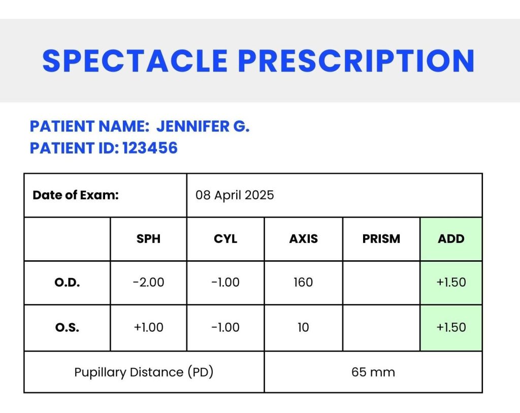
This number indicates the overall power of your lenses, measured in dioptres (D). It essentially translates the Snellen chart and visual acuity test into numerical values that indicate the strength of the lens needed for clear vision. The optical power of your lens is meant to correct refractive errors such as nearsightedness (myopia) or farsightedness (hyperopia).
If your prescription has a negative value (e.g., -2.50 D), it means you are nearsighted (myopic). Myopic eyes undergo something called ‘axial elongation’ due to a combination of molecular signals and tissue changes. The eyeball starts to become longer and the membranes are stretched backwards – contributing to higher risk of certain eye diseases in later life. This is why protective lifestyle habits and early interventions to slow myopia progression are best, and a priority for public healthcare around the world.
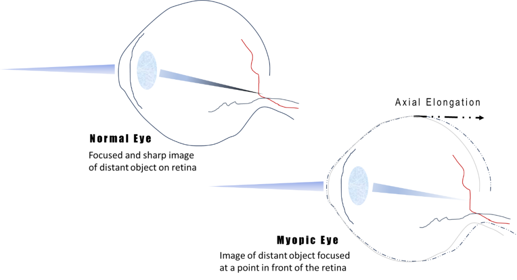
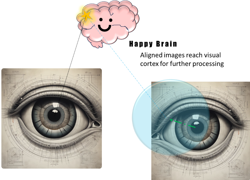
In myopia that starts in childhood and progresses, on account of axial elongation, the refractive surfaces at the front (anterior) of the eye are only able to focus images of distant objects at a point in front of the retina (posterior surface) and its image-forming cells, as seen in the image below. As a result, images of those distant objects transmitted to our brain are blurry. The higher the negative dioptre value, the more severe the myopia, requiring stronger corrective lenses worn to alter refractive power and achieve clear, focused vision.
The time to watch the dioptre value on your prescription and manage myopia and it’s risks ranges from the age of 5 to early 20s in the Singaporean cohort. Adult myopia progression is a possibility for those in intense tertiary education such as medical school, or in fields that involve a lot of near work such as long hours with microscopes.
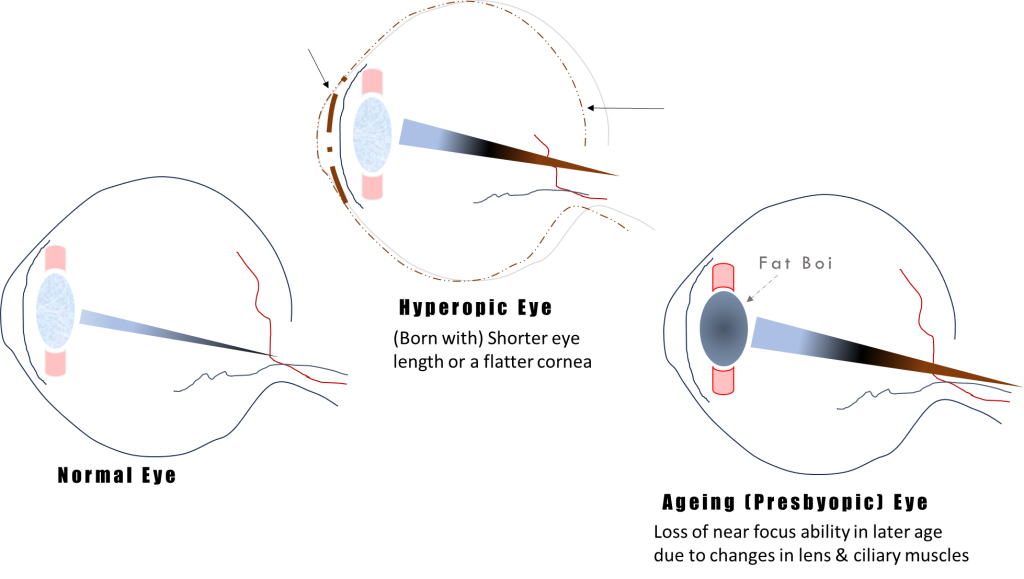
If your prescription has a positive value (e.g., +1.00 DS) it means that you are farsighted, a condition known as hyperopia. This occurs when the eye focuses images behind the retina instead of directly onto it, making close-up objects appear blurry while distant objects remain clearer. Reasons might include a cornea flatter than normal, or an eye length that is shorter than average. The higher the positive dioptre value, the more difficulty you may have seeing nearby objects, often requiring stronger corrective lenses for activities like reading or using a phone.
When farsightedness develops and worsens from around the ages of 40 to 70, this is known as presbyopia or ‘old man eye’, which is quite rude but accurate. The loss of near focus ability (accommodation) is mainly due to age-related changes of the lens and tiny muscles which control its thickness. Presbyopia is reflected in the ‘ADD’ term on your chart, explained a bit further below.
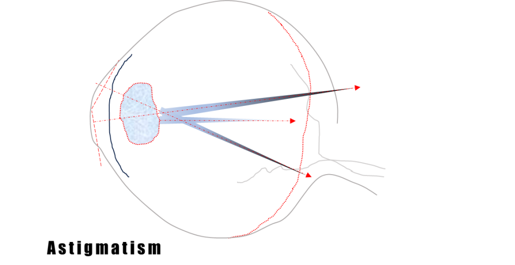
This one is a bit more complicated to understand, but the condition is quite common and requires two measurements in a prescription. The cylinder and axis values represent astigmatism, a condition where the cornea or lens has an irregular shape. Even the retina’s surface may be irregular in some astigmatism cases, but this is quite rare.
The cylinder (CYL) value in a spectacle prescription represents the difference in curvature between two principal meridians (dividing lines) – of the cornea or lens. In simpler terms, because the curved and refractive surfaces of the eye don’t ‘match’, the image ends up focused at multiple scattered points instead of a single point. Also in an astigmatic eye, the image focuses in front of and behind the retina, and this difference is the amount (cylinder value, CYL) required to shift the image to a single point on the retina. CYL is typically presented as a negative value in minus-cylinder notation, commonly used to correct myopic astigmatism, which is more prevalent. Positive-cylinder notation, indicating hyperopic astigmatism, is less commonly used in modern clinical practice.
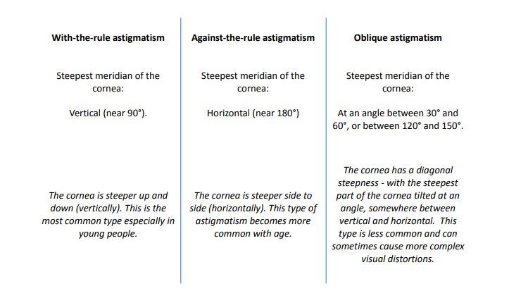
Even if two people have the same amount of astigmatism in terms of CYL, their eyes can look and behave very differently depending on the axis. Axis is defined as the orientation or direction of the cornea’s steepest or flattest curve. It is measured in degrees from 0 to 180, much like positions on a circle. The direction of the steepness changes how light bends in the eye and can affect vision in unique ways.
There are three main types of corneal astigmatism, and they are categorised based on the direction of the steepest curve, represented by the axis figure.
Each type affects how clearly you see and may require a different kind of correction in glasses or contact lenses.
The Near Addition (ADD) component in a spectacle prescription refers to the ‘additional plus power’ needed for near vision tasks, typically prescribed for presbyopic patients.
In presbyopia, the culprit is the crystalline lens, or more specifically the cells and proteins that make up the lens. This structure begins to alter in terms of chemical and mechanical properties as we age. It grows in size, changes shape, and becomes harder and less flexible so that the ciliary muscles have to work harder to control its thickness and refractive ability. Even ciliary muscles might be undergoing age-related changes, all affecting accommodation and thus the ability to clearly see near objects.
This value is added to the distance spherical power to provide appropriate magnification for reading and other close-up activities. The ADD value is the same for both eyes in most cases and usually ranges from +0.75 D to +3.00 D. This component is used in multifocal lenses such as bifocals, progressives, and reading glasses.
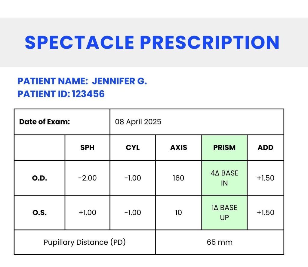
The prism component in spectacles is primarily used to correct strabismus (eye misalignment), double vision (diplopia), and other binocular vision anomalies. Prisms work by bending light before it enters the eye, compensating for alignment issues between both eyes.
Strabismus is the condition that occurs when the eyes do not align properly. One eye may look straight ahead, while the other is turned inward, outward, upward, or downward. This misalignment causes the brain to receive two different images, which can lead to confusion or double vision. Prism correction helps by shifting the image seen by the misaligned eye so both eyes can focus on the same object. This reduces the need for the brain to fuse the two separate images from each eye and helps align the visual fields.
There are also instances where no physical misalignment of the eyes is present, but prisms are still required. These situations may involve binocular vision anomalies, where the eyes appear properly aligned, but the brain struggles to coordinate the two visual inputs, leading to symptoms such as eye strain, headaches, or discomfort. In such cases, prisms help by aligning the images seen by each eye, facilitating better coordination between the two.
Prism strength is measured in prism dioptres (Δ), where higher values indicate stronger corrections. Base direction refers to the orientation of the prism, determining the direction in which the image will be shifted. The most common base directions are:
A contact lens prescription differs from a spectacle prescription due to the variation in lens positioning relative to the eye. Because contact lenses sit directly on the cornea, the prescription values may differ from those of glasses. Additionally, a contact lens prescription includes specific parameters such as base curve (BC) and diameter (DIA) to ensure a proper fit on the eye.
It is essential to consult your optometrist when obtaining a contact lens prescription, as an improper fit can affect vision and eye health. Always ensure that you have a separate, updated prescription specifically for contact lenses.
Some aspects are a bit complicated, so if you have any questions or concerns about your prescription, it is always a good idea to consult your optometrist to ensure your lenses provide optimal vision and comfort. Whether for glasses or contact lenses, regular eye exams and updated prescriptions are crucial for maintaining your eye health. This can range from at least once a year to several times a year for orthokeratology patients or the elderly that have a high risk of ocular diseases. Early detection of potential issues is key, even if you aren’t experiencing noticeable symptoms.